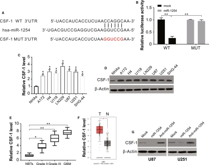
Figure 4.

Targeting relationship between miR‐1254 and CSF‐1 in glioma. (A) The predicted base pairing in miR‐1254 and CSF‐1 from TargetScan. (B) Validation of targeting relation between miR‐1254 and CSF‐1 through dual‐luciferase reporter assay. (C) The expression of CSF‐1 was higher in glioma cell lines than that in normal cell line NHAs. (D) CSF‐1 protein expression was higher in glioma cell lines than that in normal cell line NHAs. (E) Relative CSF‐1 expression in glioma samples divided according to pathological classification and NBTs. The Student's t test was used to analyse significant differences between groups. (F) TCGA database showing increased CSF‐1 expression in glioma compared with normal samples. (G) Western blotting of CSF‐1 in U87 and U251 cells transfected with indicated miR‐1254 or anti‐miR‐1254. Results were expressed as means ± SD of three independent experiments. *P < .05; **P < .01
