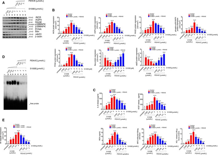
Figure 2.

Synoptic framework showing the effect of exogenous increasing concentrations of S100B proteins human control mucosal biopsies and its negative modulation by PENVE. (A) Immunoblot analysis of the immunoreactive bands showing the expression of iNOS, AQP4, RAGE, phospho‐p38MAPK, PCNA, Bax and wtp53 in control mucosal biopsies challenged with exogenous S100B protein (0.005‐5 µmol/L) given alone or at the maximum concentration (5 µmol/L) in the presence of PENVE (0.005‐5 µmol/L) at 24 h; (B) relative densitometries. In the same experimental conditions, this figure shows the quantification of (C) IL‐6, VEGF, MDA, MPO and nitrite release from the same treated mucosal biopsies. (D) EMSA analysis showing NF‐kappaB expression and (E) its relative densitometric quantification in the same experimental conditions. Results are expressed as mean ± SEM n = 5 experiments in triplicate; ***P < .001, **P < .01 and *P < .05 vs unstimulated control group and °°°P < .001, °°P < .01 and °P < .05 respectively, vs S100B 5 µmol/L group
