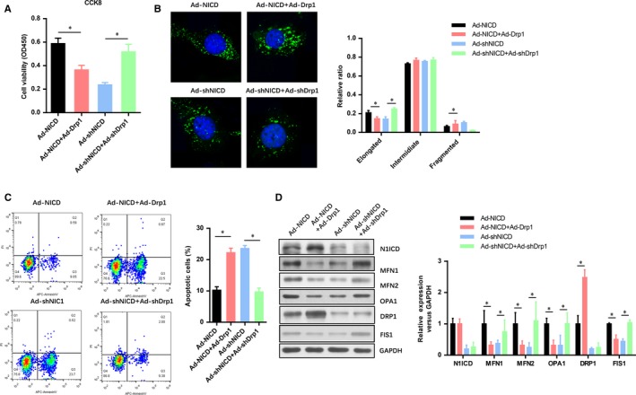
Figure 3.

DRP1 is critical to the Notch1 improved cell viability and mitochondrial fusion in myocardiocytes exposed to IRI. A, The myocardiocytes infected with indicated adenovirus were exposed to H/R and evaluated by CCK‐8 assays. B, After H/R, confocal microscopy analysis of the mitochondria fusion and fission in myocardiocytes infected with indicated adenovirus. C, FACs analysis the apoptotic cell numbers of myocardiocytes infected with indicated adenovirus. D, The level of N1ICD and mitochondrial fusion‐fission markers (MFN1, MFN2, OPA1, DRP1, AND FIS1) were evaluated by Western blot. All data are means ± SEM *P < .05 with the indicated group
