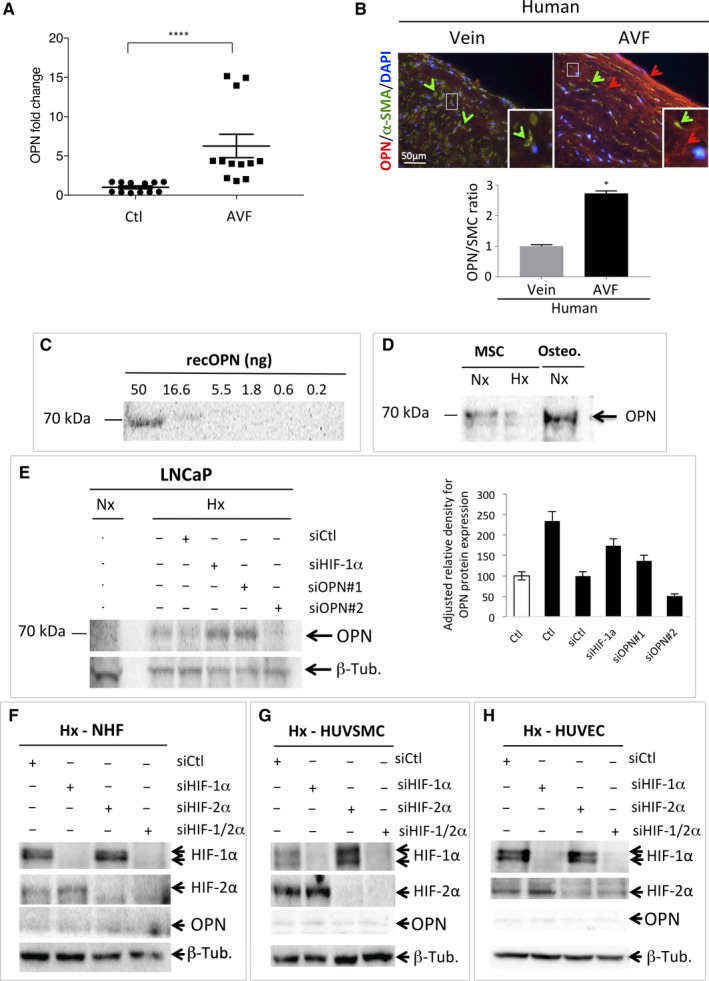
Figure 1.

Osteopontin (OPN) is weakly expressed in NHF, HUVSMC and HUVEC and is not induced in hypoxia. A, Levels of OPN mRNA in AVF patients (n = 12) compared to control veins (Ctl). ****, P < .0001 (Test Mann‐Whitney). B, Representative immunofluorescence of OPN (red staining and arrow) and alpha‐smooth muscle actin (α‐SMA—green staining and arrows) of human control upper‐arm vein and venous limb of a mature AVF. Osteopontin quantification of group data (vein, n = 3; AVF, n = 2), unpaired t test, P < .01 (right). C, Quantitative immunoblotting to detect recombinant OPN protein. Serial dilutions from 50 to 0.2 ng were used. D, Bone marrow mesenchymal (MSC) and osteoblast (Osteo.) cell lysates analysed by immunoblotting for OPN as indicated. E, LNCap cells were transfected with control siRNA (siCtl), siHIF‐1α (40 nmol/L), siOPN#1 (40 nmol/L) and siOPN#2 (40 nmol/L) and incubated in normoxia (Nx) or hypoxia 1% O2 (Hx) for 48 h. Cell lysates were analysed by immunoblotting for OPN or β‐tubulin as indicated. Quantification of the signal to OPN is shown using ImageJ (OPN/β‐tubulin). F–H, NHF (F), HUVSMC (G) and HUVEC (H) cells were transfected with control siRNA (siCtl), siHIF‐1α (40 nmol/L), siHIF‐2α (40 nmol/L) and siHIF1/2α (40 nmol/L + 40 nmol/L), incubated in hypoxia 1% O2 (Hx) for 48 h. AVF, Arteriovenous fistula; HUVEC, human umbilical vein endothelial cells; HUVSMC, Human umbilical vein smooth muscle cells; NHF, normal human fibroblasts
