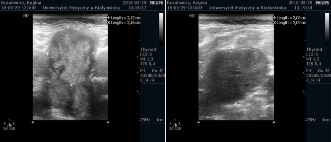
Figure 2.
Neck usg. (A) The cross-section showed thyroid nodule with a diameter of 21 × 22 mm in the middle of the left lobe with irregular outline; (B) showed conglomerates of hypoechoic cervical masses without echogenic hilus suspicious for enlarged LNs (31 × 21 × 30 mm in size) on the right side.

 This work is licensed under a