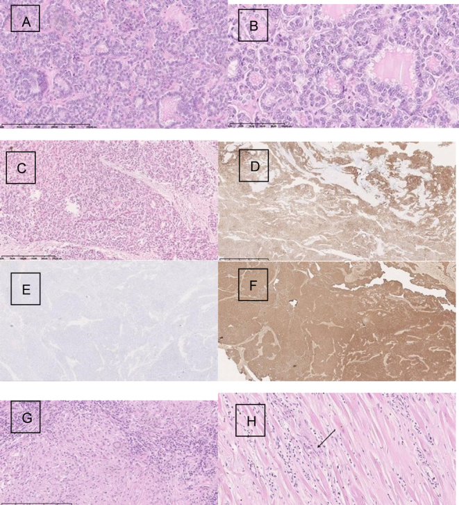
Figure 3.

Histopathology of pituitary metastasis at diagnosis. (A) Thyroid gland papillary carcinoma follicular variant. Magn. 100×; (B) Papillary cancer of thyroid gland-cytological features confirm the recognition. Magn.200×; (C) Thyroid gland papillary cancer metastasis into the sellar region. Magn. 40×; (D) Cytokeratin 7 expression within the tumor cells. Magn. 40×; (E) Cytokeratin 19 negative expression within the metastase. Magn. 40×; (F) Thyreoglobulin expression within the cancer cells in metastasis. Magn. 40×; (G) Granuloma with giant cells, macrophages, and lymphocytes within the lymph node. Magn.200×; (H) Granuloma within the cardiac muscles. Magn. 40×.

 This work is licensed under a