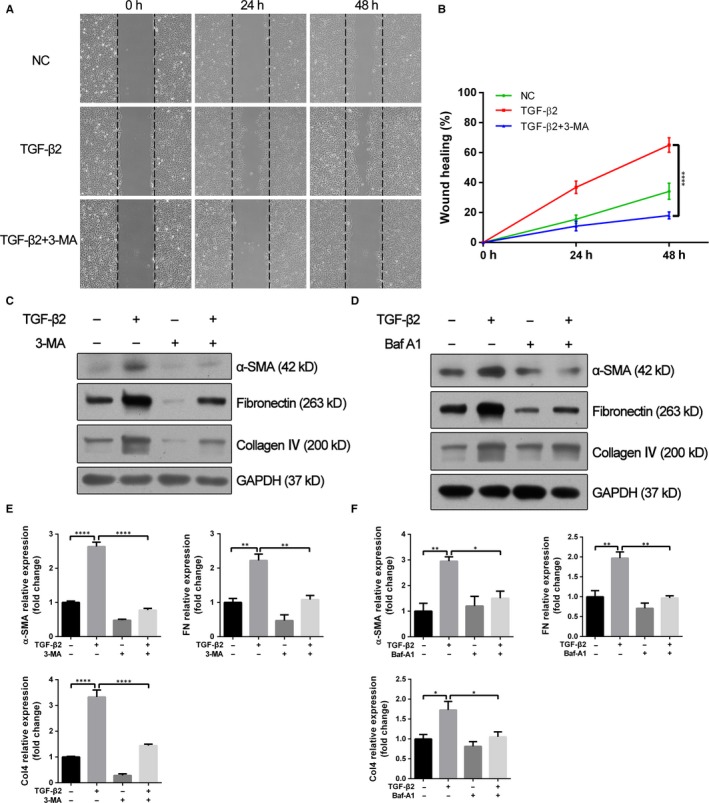
Figure 3.

Autophagy inhibitors attenuate TGF‐β2–induced cell migration and EMT markers synthesis. A, Wound healing assays of TGF‐β2 (10 ng/mL) stimulated ARPE‐19 cells co‐treatment with or without 3‐MA (10 mmol/L). Phase‐contrast microphotographs (4 × objective) were acquired at 0, 24 and 48 h after scratching. B, Graph shows the percentage of wound healing area relative to 0 h. The data are presented as mean ± SEM, n = three independent experiments. ****P < .0001. C, D, Western blot analysis of α‐SMA, fibronectin and collagen IV in ARPE‐19 cells treated with TGF‐β2 (10 ng/mL) in the absence or presence of either 3‐MA (10 mmol/L) or Baf‐A1 (10 nmol/L) for 24 h. GAPDH was used as loading control. E, F, Bar graphs indicate the relative expression levels of α‐SMA, fibronectin and collagen IV (normalized to GAPDH) in Western blot analysis. The data are presented as mean ± SEM, n = three independent experiments. *P < .05, **P < .01 and ****P < .0001
