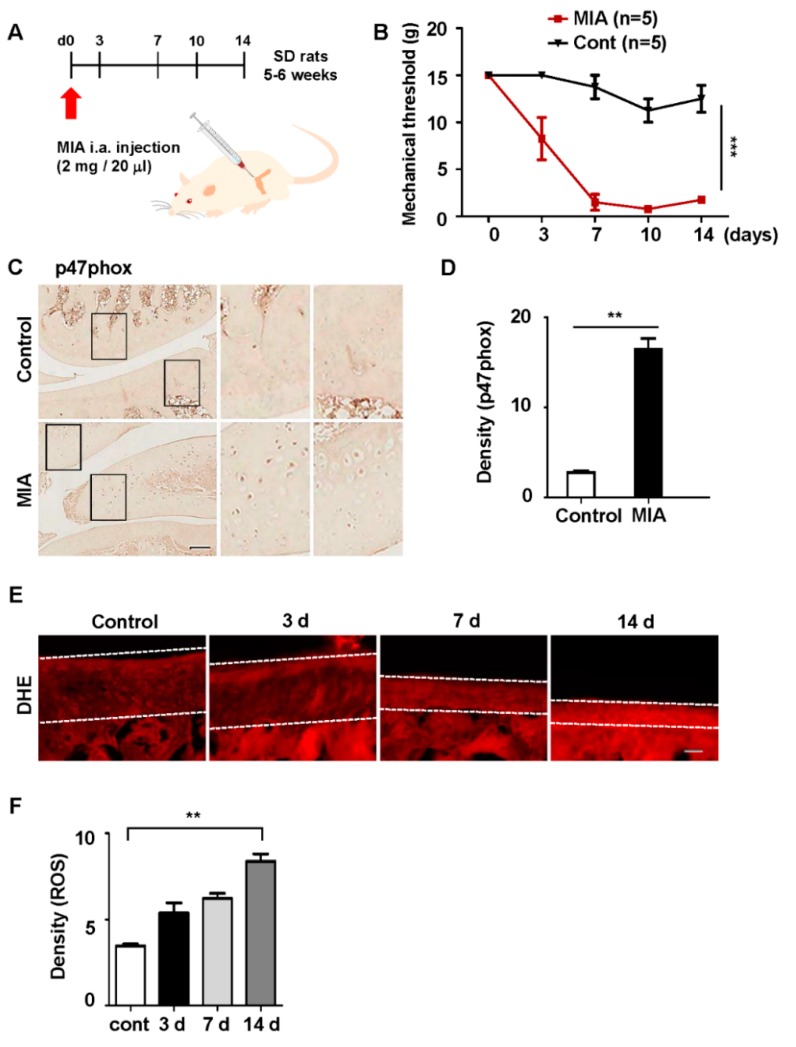
Figure 2.
Monosodium iodoacetate (MIA)-induced expression of p47phox and the production of cellular ROS. (A) Prior to MIA injection, the rats were subjected to a von Frey filament test; only those that met a predefined threshold were selected for MIA injections. (B) The von Frey test was repeated on days 3, 7, 10, and 14 after injection; all data are presented as the mean ± standard error of the mean. (C) Rat knee tissues were immunostained with an anti-p47phox antibody at 3 days after MIA injection; scale bar = 50 µm. (D) The density of p47phox expression in the knee was measured by using ImageJ software; all data are presented as the mean ± standard error of the mean. (E) DHE fluorescence imaging of the knee in the OA rat model; white lines indicate cartilage in the tissue. (F) Quantification of DHE fluorescence; all data represent the mean ± standard error of the mean (error bars) of three experiments.

