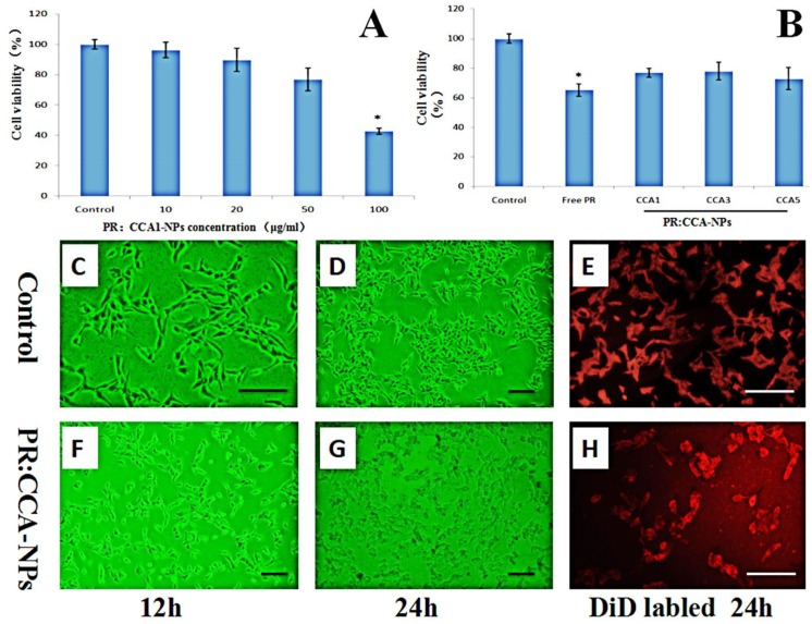
Figure 5.
Effects of different concentrations of nanoparticles (A) and different PR:CCA-NPs (B) on the proliferation ability of B16 cells (24 h); Observing the B16 cells of mice at different time under inverted microscope (C, D, F, G); Cells were stained with DiD to visualize membrane morphology (E, H). Note: the scale bar represents 25 µm; * p < 0.05 vs control.

