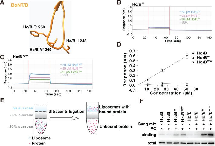
Fig 3. Introducing aromatic residues at the tip of HCB-LBL enables its binding to lipid membranes.
(A) Structure of HCB-LBL, with three residues at the tip marked. (B–C) Binding of HC/B mutants containing either a single point mutation, I1248W, or double point mutations, I1248W/V1249W, to ND1 were analyzed by BLI assays. HC/BW showed low-level binding to ND1, whereas HC/BWW showed robust binding. (D) The maximal binding signals of HC/B mutants as measured in panels B and C were averaged and plotted (mean ± SD, n = 3). Numerical values for (D) are available in S1 Data. (E–F) Binding of WT HC/B, HC/BW, and HC/BWW to liposomes containing POPC or POPC plus gangliosides were analyzed by liposome flotation assays. Proteins bound to liposomes floated to the top of the sucrose density gradient and were collected and subjected to immunoblot analysis. HC/BW and HC/BWW were able to bind POPC liposomes, and the presence of gangliosides further enhanced their binding. BLI, biolayer interferometry; BoNT, botulinum neurotoxin; BSA, bovine serum albumin; HC, C-terminal receptor-binding domain; LBL, lipid-binding loop; ND1, receptor-free nanodisc; PC, phosphatidylcholine; POPC, 1-palmitoyl-2-oleoyl-glycero-3-phosphocholine; WT, wild-type.

