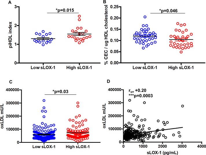
Fig 3. Assessment of lipid function in low and high sLOX-1 SLE patients.
(A) Increased proinflammatory HDL (piHDL) in high sLOX-1 (n = 20) SLE patients versus low sLOX-1 (n = 16) SLE patients using DCFDA as a substrate to measure LDL oxidation; (B) Cholesterol efflux capacity of SLE HDL from donors with low sLOX-1 (n = 36) and high sLOX-1 (n = 33). Global cholesterol efflux capacity (CEC) was normalized to total HDL cholesterol quantification from each donor before being matched with sLOX-1 levels. (C) Levels of oxLDL in high (72902 ± 5345 mU/L, n = 93) and low sLOX-1 SLE patients (59489 ± 3396 mU/L, n = 163). (D) Spearman correlation (rs) between oxLDL and sLOX-1. Data represented as mean ± SEM. * p<0.05, ** p<0.01, ***p<0.001 and ****p<0.0001.

