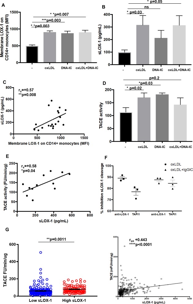
Fig 5. Relationship between induction of membrane LOX-1/sLOX-1 cleavage with TACE activity.
Healthy monocytes were isolated from PBMCs and treated with oxLDL (30 μg/mL) for 3h followed by DNA-IC (20 μg/mL) (co)stimulation for 24h. (A) LOX-1 (MFI) expression on CD14+ monocytes. (B) sLOX-1 (pg/mL) detection from supernatant post 24h treatment. (C) Spearman correlation (rs) between membrane LOX-1 (MFI) and sLOX-1 (pg/mL). (D) Tumor necrosis factor alpha activating enzyme (TACE) activity (FU/min/μg) was measured using a fluorogenic peptide substrate (Mca-PLAQAV-Dpa-RSSSR-NH2) from supernatants post 24h treatment. (E) Spearman correlation (rs) between sLOX-1 (pg/mL) and TACE activity (FU/min/μg). Data from in vitro experiments represented as mean ± SEM. (F) Inhibition of sLOX-1 cleavage in the presence of either the TAPI-1 inhibitor (100 μM) or the LOX-1 receptor blocking antibody. (G) TACE activity measurements normalized to total protein in SLE patients with low (2.741 ± 0.3491, n = 168) and high (5.134 ± 0.7696, n = 100) sLOX-1. Relationship between TACE activity (FU/min/μg) and sLOX-1 (pg/mL) from matched patient serum shown by Spearman correlation (rs). Data represented as mean ± SEM. * p<0.05, ** p<0.01, *** p<0.001 and **** p<0.0001.

