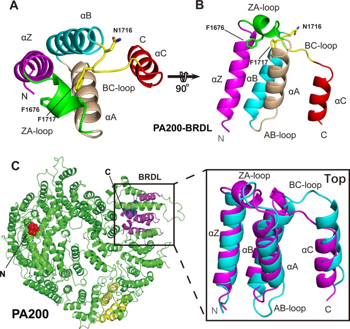
Fig 7. The BRDL domain of PA200.
(A-B) Ribbon representation of the PA200 BRDL domain in top view (A) and side view (B), with the key residues shown as sticks. (C) The alignment of the PA200 BRDL domain (magenta) and the corresponding region (cyan) of Blm10. The region corresponding to BRDL domain of Blm10 is shown as yellow. The N-terminal of Blm10 is shown as red spheres, whereas the C-terminal is indicated as blue spheres (see also S4 Fig). Blm10, Bleomycin resistance 10; BRDL, bromodomain-like; PA200, proteasome activator 200.

