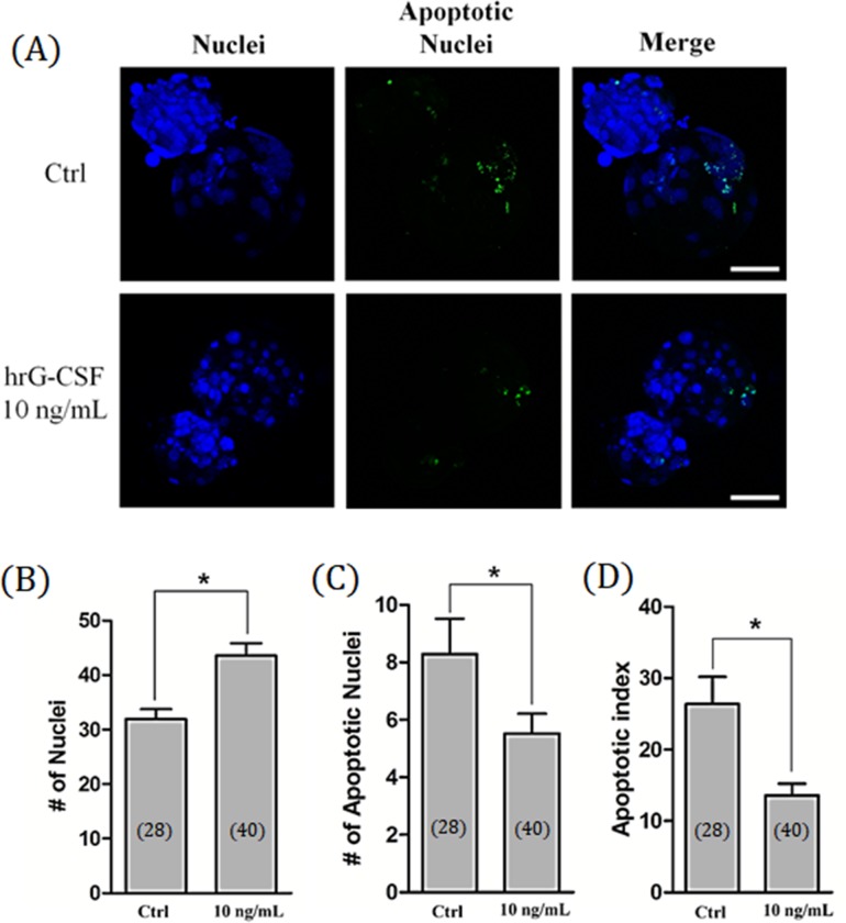
Fig 2. Total cell number and apoptosis of cloned blastocyst after hrG-CSF treatment.
Representative laser scanning confocal microscopy images (400×) of nuclei (blue) and fragmented DNA (green) in porcine blastocysts after culturing for 7 days with (10 ng/mL) or without (Control) hrG-CSF treatment. Scale bar = 100 μm (A). The total cell number of nuclei (B), apoptotic nuclei (C), and apoptotic index (D) in porcine cloned blastocysts developed in vitro. Ctrl: no treatment; hrG-CSF 10 ng/mL: human recombinant granulocyte-colony stimulating factor 10 ng/mL treatment for entire stage (Days 0 to 7). The number of embryos examined in each experimental group is shown in parentheses. *: P < 0.05.

