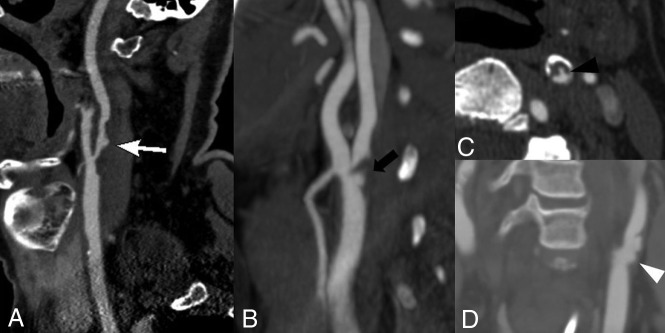
Fig 3.
Plaque surface morphology and ulceration. A, A 69-year-old woman with a large soft plaque with an irregular surface and focal plaque ulceration (white arrow) with the plaque narrowing the proximal left ICA. B, A 62-year-old man with focal soft plaque at the right carotid bifurcation and proximal ICA with a large plaque ulceration (black arrow). C, A 73-year-old man with a predominant soft plaque with peripheral calcification narrowing the left ICA with focal plaque ulceration (black arrowhead) extending into the soft plaque. D, A 67-year-old woman with irregular, ulcerated plaque (white arrowhead) best seen on the coronal reconstruction. There is no significant associated luminal narrowing with this irregular, ulcerated plaque.

