Fig 1.
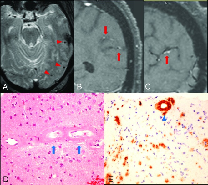
Arterial wall enhancement is associated with amyloid accumulation within the vessel wall. A, Gradient-echo image shows microhemorrhages in the left temporal lobe (red arrowheads). B and C, coronal and sagittal views of postcontrast T1-weighted VWMRI show enhancement in the wall of cortical branches of the left middle cerebral artery in the parietal and temporal lobes (red arrows). D, Hematoxylin-eosin stain of a left temporal lobe sample shows thickened, hyalinized blood vessels containing amorphous eosinophilic material (blue arrows) in small- and medium-sized arteries within the leptomeninges and superficial cortical gray matter. E, Immunostain for β-amyloid shows amyloid accumulation within the vessel wall (blue arrowhead). No inflammatory cells were observed surrounding the vessels.

