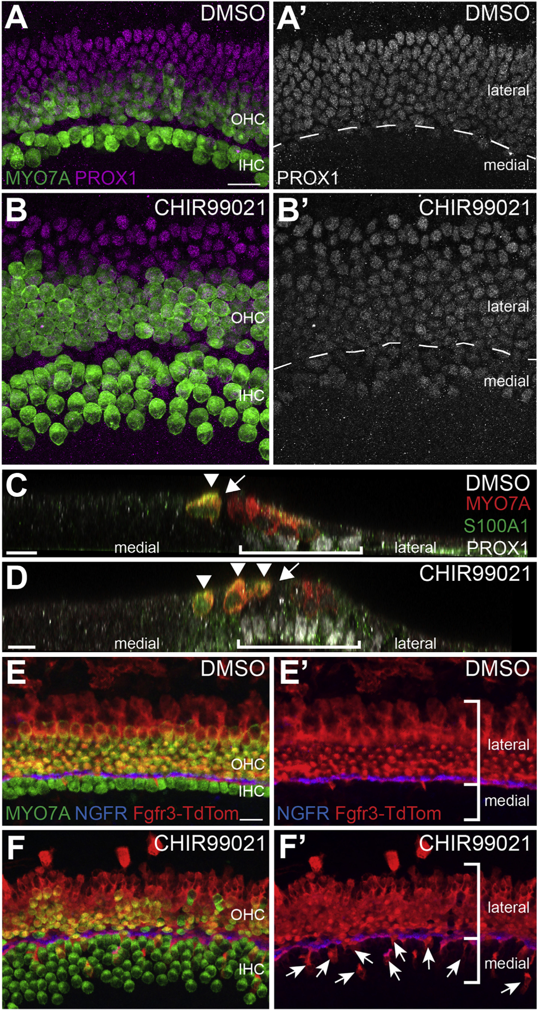
Fig. 6. Disruption of the medio-lateral boundary in GSK3-inhibited explants is partly due to lateral cells adopting medial cell fates.

A, B. Control (A, A’) and CHIR-treated (B, B’) explants labeled with anti-MYO7A (green) and anti-PROX1 (magenta). In DMSO-treated controls the boundary of PROX1 aligns precisely with the row of IPCs (dashed line in A’). In contrast, in CHIR-treated explants, PROX1 expression extends into the medial domain (below dashed line in B’). C, D. Confocal orthogonal views of the sensory epithelium from control (C) or CHIR-treated explants (D). Hair cells are labeled in red (MYO7A) and PROX1+ supporting cells are in white. S100A1 in green indicates IHCs and Deiters’ cells. In the control, PROX1+ cells (bracket) are located lateral to the pillar cells (arrow). In contrast, in the CHIR-treated explant PROX1+ supporting cells are present beneath extra IHCs (D, arrowheads). Pillar cells are indicated with an arrow. E-F’. Cochlear explants from transgenic Fgfr3creErt2;R26RTdTom mice were established at E13.5 and maintained in either DMSO or CHIR from E13.5 – E17.5. Explants were treated with 1 μM 4-OH-Tamoxifen from E13.5 – E15.5 to induce recombination. E, E’. In controls, Fgfr3creErt2;R26RTdTom positive cells (red) are largely restricted to the lateral domain. Hair cells are labeled with MYO7A (green) and IPCs are labeled with NGFR (blue). (F, F′) Inhibition of GSK3 causes a significantly increased number of lateral, Fgfr3creErt2;R26RTdTom positive cells to adopt medial IHC, inner phalangeal cell, or border cell fates. Labeling as in E. Arrows indicate medial cells that are tdTomato-positive. Scale bars = 20 μm.
