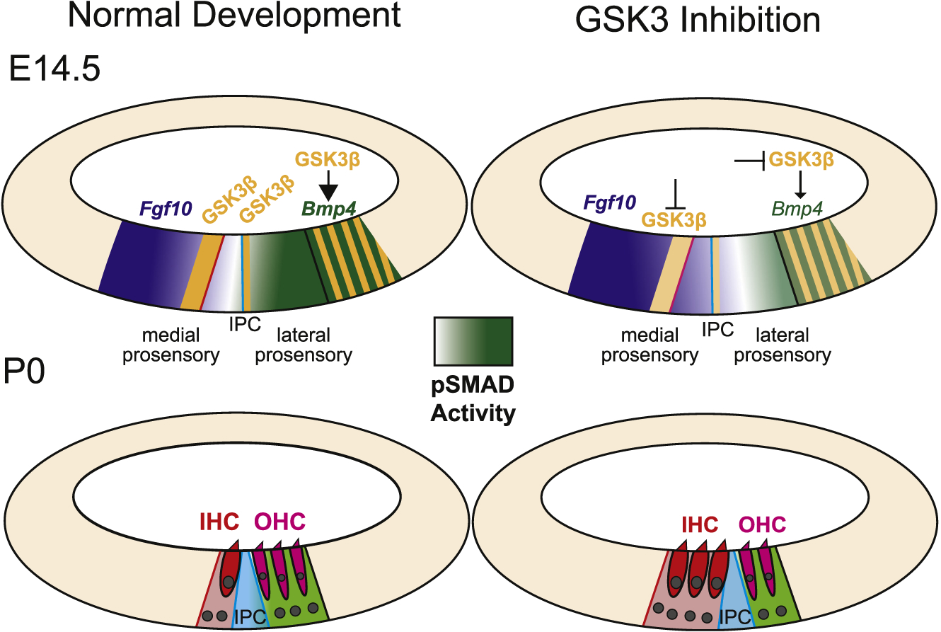
Fig. 9. Summary of the effects of inhibition of GSK3.

At E14.5 the cochlear duct contains medial and lateral prosensory domains. GSK3β (orange) is expressed at the boundaries between each of the two domains and between the prosensory and non-sensory cells. The boundary between medial and lateral prosensory domains (blue line) aligns with the position of the inner pillar cell (IPC). At the lateral boundary, GSK3β expression overlaps with Bmp4. Data presented in this study illustrates that Bmp4 expression is dependent on GSK3 activity. Expression of Bmp4 leads to a gradient of pSMAD that extends medially (light green). The level of pSMAD acts to maintain the boundary between the medial and lateral prosensory domains. As a result, at P0, cells within the medial prosensory domain have developed as inner hair cells (red) while cells in the lateral prosensory domain have developed as outer hair cells (magenta). When GSK3 activity is inhibited, two changes occur. Bmp4 expression decreases, leading to a lateral shift in the gradient of pSMAD. As a result, the lateral prosensory domain is decreased in size, leading to a relative change in the position of the IPC, more IHCs, and fewer OHCs. In addition, the medial boundary between prosensory and non-sensory regions is shifted medially, leading to an additional increase in IHCs. The molecular basis for the medial expansion of the medial prosensory domain is not clear but may be mediated through changes in Notch pathway activity.
