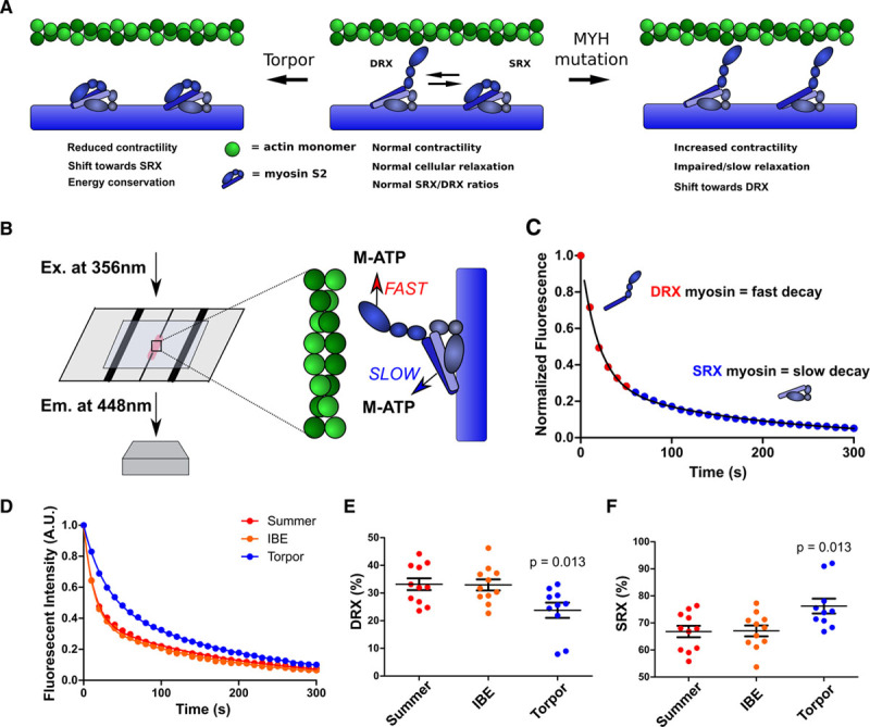
Figure 1.

Mant-ATP assays assess physiological and pathological changes in cardiac myosin conformations. A, Depiction of the predicted myosin SRX and DRX conformations, indicating the dynamic effects of the interacting heads motif, in torpor (Left), normal cardiac function, and in hypertrophic cardiomyopathy (Right). B, Schematics depicting Mant-ATP (M-ATP) infused into a flow chamber containing permeabilized cardiac tissue. A subsequent chase of dark ATP results in fast and slow fluorescence decay (detected at 448 nm using a 40× objective) from DRX and SRX conformations of myosins, respectively. C, Representative Mant-ATP fluorescent chase experiment in wild-type tissues, described by a double-exponential decay. D, Mant-ATP assays of myocardium from ground squirrel hearts, obtained during summer arousal, interbout euthermia (IBE), and torpor. E and F, Proportions of myosin heads in the DRX conformation (E) with higher rates of ATP cycling and in SRX conformation (F) with slower ATP cycling. Data were obtained from studies of 3 hearts in each physiological state, with 3 to 4 samples studied per heart and are plotted as mean±SEM, with significance tested in comparison with summer (arousal), by 1-way ANOVA with Bonferroni correction with a significance cutoff at P<0.05. A.U. indicates arbitrary units; DRX, disordered relaxed state conformation of myosin molecule; Em., emission; Ex., excitation; and SRX, super relaxed state conformation of myosin molecule.
