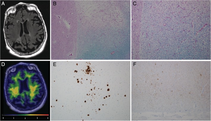
Figure 4.

Imaging and pathologic evaluation of a multiple sclerosis (MS) patient, a female 81‐year‐old APOE ε4 carrier. (A) Brain magnetic resonance imaging showed generalized cerebral and cerebellar atrophy and extensive white matter changes with T2 hyperintensities in the white matter, particularly in the periventricular regions. (B, C) Anterior basal ganglia (level of head of caudate nucleus) with adjacent white matter chronic demyelinating lesion (luxol fast blue/periodic acid Schiff). Original magnification, (B) × 40 and (C) × 100. (D) Pittsburgh compound B (PiB) positron emission tomography (PET) was negative with an Alzheimer disease signature PiB SUVr of 1.39. (E) Middle frontal gyrus with sparse neuritic plaques and moderate diffuse plaques (β‐amyloid immunostain). (F) Tau immunostain of entorhinal cortex with sparse neurofibrillary tangles. (E, F) Original magnification, ×100.
