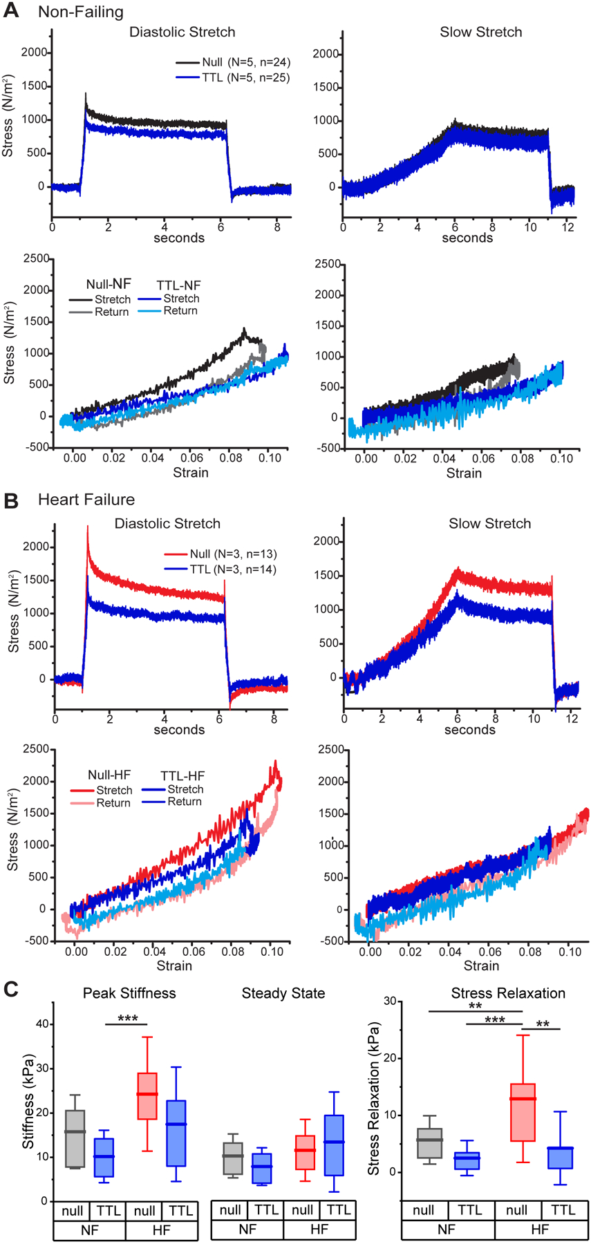
Figure 3: Overexpression of TTL is sufficient to reduce viscoelasticity in failing and non-failing human myocytes.

A-B, Average diastolic (left) and slow (right) stress-time (top) and stress-strain (bottom) plots in intact myocytes from NF and HF hearts following culture with either null-encoding adenovirus (Black, Red) or adenoviral overexpression (OE) of TTL (Blue). C, Diastolic and steady state stiffnesses (left) and stress relaxation (right) in NF and HF cardiomyocytes infected with null or TTL OE adenovirus. Statistical significance determined by two-way ANOVA with factors of etiology (NF or HF) and treatment (TTL OE vs. null). All significant interactions from this analysis are denoted in C, while significant factor effects are stated in the text, but not denoted on the figure for simplicity. Significance denoted as **p<0.01 and ***p<0.001
