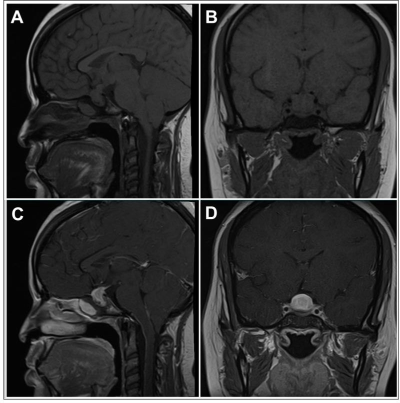
Figure 4: Magnetic Resonance Imaging Findings in a Case of Primary Hypophysitis.
Panel A) T1-weighted image, sagittal section. Panel B) T1-weighted image, coronal section. Panel C) T1-weighted image post-gadolinium, sagittal section. Panel D) T1-weighted image post-gadolinium, coronal section. A homogeneous enlargement of the pituitary with thickening of the stalk can be seen. The mass shows intense and homogeneous enhancement post-gadolinium.
With permission from ref. 46
Prete A, Salvatori R. Hypophysitis. in: Feingold KR1, Anawalt B2, Boyce A3, Chrousos G4, Dungan K5, Grossman A6, Hershman JM7, Kaltsas G8, Koch C9, Kopp P10, Korbonits M11, McLachlan R12, Morley JE13, New M14, Perreault L15, Purnell J16, Rebar R17, Singer F18, Trence DL19, Vinik A20, Wilson DP21, editors. Endotext [Internet]. South Dartmouth (MA): MDText.com, Inc.; 2000-.
2018 Aug 15.

