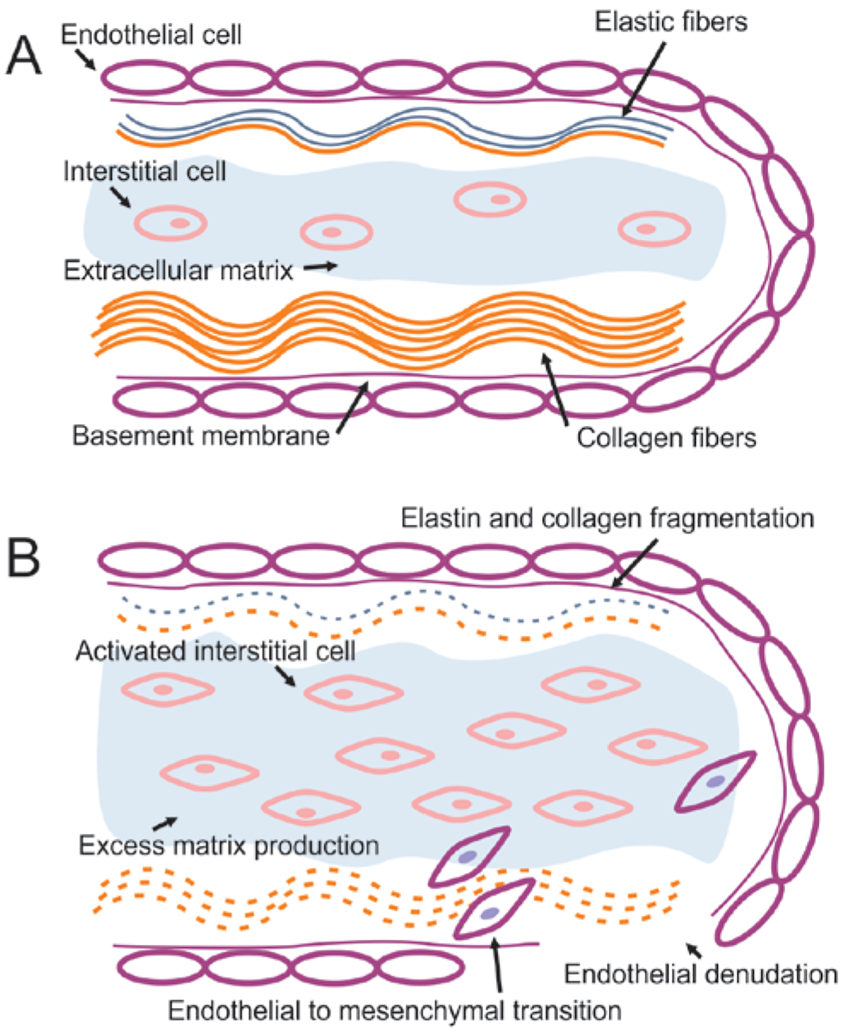
Figure 1.

Normal mitral valve structure (A) and mechanism of myxomatous mitral valve disease (MMVD) (B). The normal valve is made up of a layer of endothelial cells surrounding the atrialis layer, which is made up of elastic and collagen fibers, the spongiosa layer, which consists of extracellular matrix (ECM) rich in proteoglycans and the occasional valvular interstitial cell, and the fibrosa, which consists of tightly packed collagen fibers. In valves affected by MMVD, the interstitial cells are activated into a myofibroblast-like phenotype, which is accompanied by excessive deposition of ECM, dissolution and fragmentation of the elastic and collagen fibers of the atrialis and fibrosa, endothelial to mesenchymal cell transformation and migration of endothelial cells into the spongiosa, and denudation of the endothelial cell lining and exposure of the subendothelial collagen.
