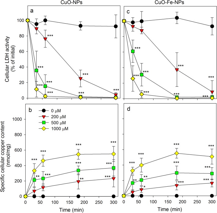
Fig. 4.
Consequences of an application of iron-free CuO-NPs and CuO-Fe-NPs on the viability and the copper content of C6 glioma cells. The cells were treated without or with CuO-NPs (a, b) or CuO-Fe-NPs (c, d) dispersed in the concentrations indicated in IB-BSA for up to 5 h at 37 °C before the cellular LDH activity (a, c) and the cellular copper content (b, d) were determined. The data shown represent means ± SD of values obtained in five independent experiments. Asterisks indicate the significance of differences of data compared with those of control cells (incubated in the absence of NPs) (*p < 0.05, **p < 0.01, ***p < 0.001; ANOVA)

