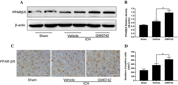
Fig. 7.
Pretreatment with GW0742 increased the PPAR-β/δ levels in ICH lesions on day 3. a Western blotting for PPAR-β/δ, assessed using perihematomal tissue on day 3. b The optical density of PPAR-β/δ relative to β-actin in the different groups is illustrated by the quantification graph. c Representative photographs of PPAR-β/δ immunostaining in the perihematomal area on day 3 (bar = 50 μm). d The positive cells shown in C were counted. Values are mean ± SD; *P < 0.05 vs. vehicle group (n = 5/group)

