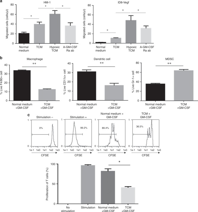
Fig. 5. The impact of GM-CSF signals on MDSC migration and differentiation.
a The number of migrated Gr-1 + cells in response to HM-1 or ID8-Vegf cell-conditioned medium cultured under normoxic or hypoxic conditions. Gr-1 + cells were treated with rat IgG control or anti-GM-CSFRa antibody *P < 0.05. Data are represented as mean ± SEM, n = 5 per group. b Flow cytometric analysis for the presence of TAM, DC and MDSCs induced by in vitro generation assay. CD11b + cells were cultured in the presence of GM-CSF with or without ID8-Vegf cell conditioned medium. **P < 0.005. Data are represented as mean ± SEM, n = 5 per group. c The proportions of proliferation of CD8 + T cells after co-culture with in vitro-induced myeloid cells. The histogram shows the percentages of proliferated T cells. *P < 0.05. Data are represented as mean ± SEM. n = 4 per group.

