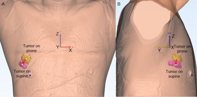
Figure 5.
A case of tumor movement at the nipple origin from prone to supine positions. (A) and (B) Show that the tumor located in the outer area in the prone position moved outward and downward along the z-axis. (A) Presents a frontal view of the 3D body surface and the nipple origin (blue point) in supine position, and two tumors from the prone position (yellow) and supine position (pink). (B) Is a right-side view. (Yellow points: the origins of tumors; red arrow: tumor movement from prone to supine positions).

