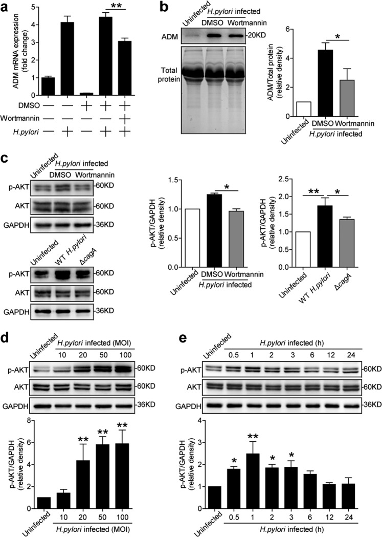
Fig. 3. H. pylori stimulates gastric epithelial cells to express ADM via PI3K–AKT pathway.
a, b AGS cells were pretreated with Wortmannin (a PI3K–AKT inhibitor) and then stimulated with WT H. pylori (MOI = 100) for 24 h. ADM mRNA expression (a) and ADM protein (b) in/from AGS cells were analyzed by real-time PCR and western blot and statistically analyzed (n = 3). The results are representative of three independent experiments. c AGS cells were pretreated with Wortmannin (a PI3K–AKT inhibitor) and then stimulated with WT H. pylori (MOI = 100) for 1 h, or were infected with WT H. pylori or ΔcagA (MOI = 100) for 1 h. AKT and p-AKT proteins were analyzed by western blot and statistically analyzed (n = 3). The results are representative of three independent experiments. d, e AGS cells were infected with WT H. pylori with different MOI (1 h) (d) or at different time points (MOI = 100) (e). AKT and p-AKT proteins were analyzed by western blot and statistically analyzed (n = 3). The results are representative of three independent experiments. *P < 0.05, and **P < 0.01 for groups connected by horizontal lines compared, or compared with uninfected cells.

