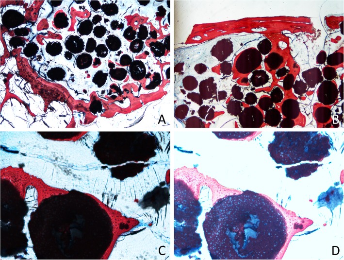
Fig. 4.
Photomicrographs of ground sections after 4 months of healing. a Bone formed from the base of the sinus. b Bone plate connected by bridges of the new bone to the close-to-window region. c Particle of the graft surrounded by new bone. d Overexposed image to show the new bone ingrowth within the granules of biomaterial

