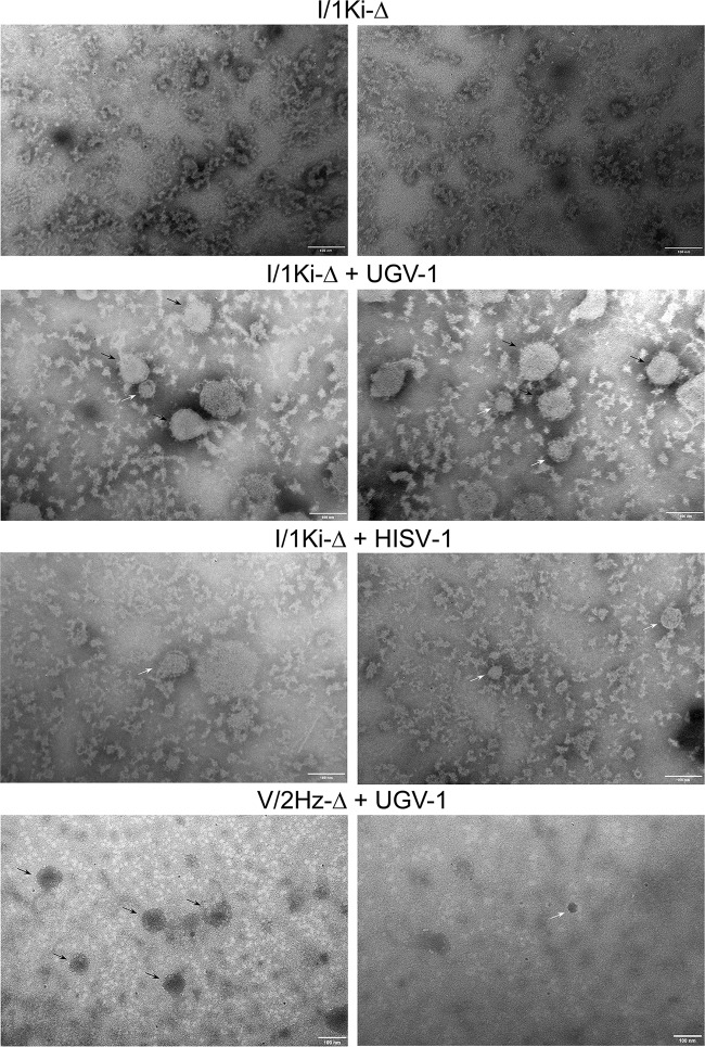
FIG 7.
Transmission electron microscopy (TEM) of pelleted supernatants. Persistently SDeV-infected I/1Ki-Δ cells were inoculated with medium collected from clean I/1Ki cells (mock) or superinfected with UGV-1 or HISV-1. V/2Hz-Δ cells were superinfected either with UGV-1 or HISV-1. The cell culture medium was collected at 2- to 3-day intervals until 14 days postinfection, after which the supernatants were pooled and filtered, followed by ultracentrifugation to pellet the virus particles. After resuspending the pelleted material, an aliquot of the pelleted material was prepared for TEM with negative staining. The top panels show the material pelleted from mock-infected I/1Ki-Δ cells, the second row of panels show the material pelleted from UGV-1-infected I/1Ki-Δ cells, the third row of panels show the material pelleted from HISV-1-infected I/1Ki-Δ cells, and the bottom panels show the material pelleted from UGV-1-infected V/2Hz-Δ cells. The black arrows in the figure panels point to UGV-1 particles, and the white arrows show putative SDeV particles as judged by size. The images were taken by using a JEOL JEM-1400 transmission electron microscope at ×200,000 magnification.

