FIG 4.
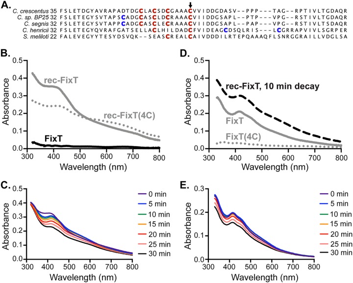
FixT binds an Fe-S cluster. (A) Sequence alignment of FixT proteins from several Caulobacter species and S. meliloti. The position of the first amino acid residue of each sequence is on the left. Cys residues aligning with the C. crescentus 4-Cys motif are in red. Additional nearby Cys residues are in blue. The position corresponding to C. crescentus C64 is marked by an arrow. (B) Absorption spectra of aerobically purified FixT (FixT), anaerobically reconstituted wild-type FixT (rec-FixT), and anaerobically reconstituted FixT(4C) [rec-FixT(4C)]. (C) Absorption spectra of rec-FixT collected at 5-min intervals after exposure to ambient air. (D) Absorption spectra of anaerobically purified H6-Sumo-FixT and H6-Sumo-FixT(4C) at 400 μM. The 10-min decay curve of FixT with a reconstituted Fe-S cluster (rec-FixT) from panel C is shown for comparison. (E) Absorption spectra of anaerobically purified H6-Sumo-FixT collected at 5-min intervals after exposure to ambient air.

