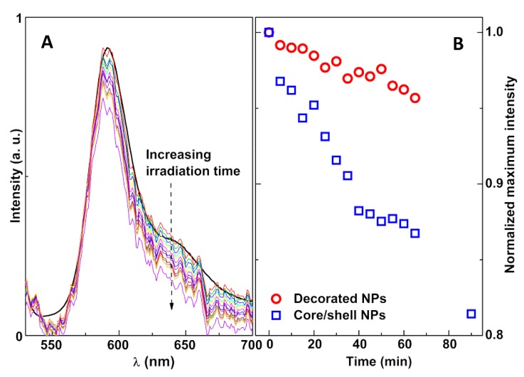
Figure 14.
(A) Fluorescence spectra of Rhodamine B-DOPE in magnetoliposomes containing nickel ferrite/gold core/shell nanoparticles following several irradiation times (5-min steps, maximum: 90 min). The black line curve is the reference spectrum obtained in a commercial fluorimeter. (B) Variation of the normalized maximum intensity of Rhodamine B fluorescence with irradiation time, for magnetoliposomes containing both types of magnetic/plasmonic nanoparticles.

