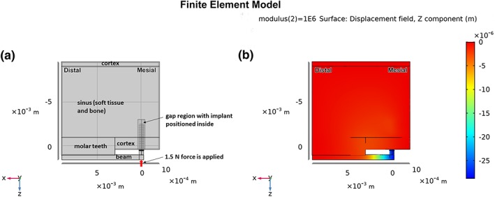
Figure 2.

(a) Overall formulation of the FE model, showing idealized regions of molar teeth, cortical bone, sinus, titanium beam, gap, and implant. The thickness of the model is 1.5 mm into the page, with mesio‐distal and occluso‐apical dimensions as noted in the text. The mesial, distal, and superior surfaces of the model are fixed; (b) semi‐transparent view of a portion of the model showing z‐displacements after loading the end of the beam with 1.5 N, and with the modulus of the gap tissue equal to 1 MPa
