. 2020 Mar 3;17(2):131–139. doi: 10.21873/cgp.20173

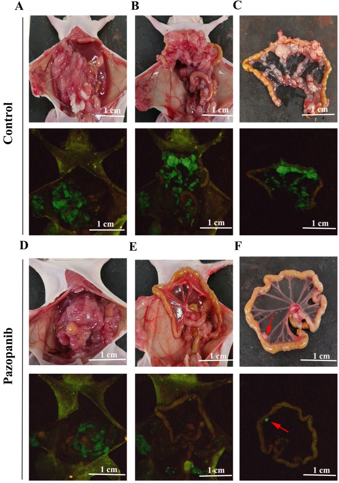
Figure 4. Photographs and fluorescence images of orthotopic CT-26-GFP colorectal cancer nude-mouse model at autopsy in the control and PAZ groups. (A-C) Representative photographs of the control group (upper panels) and corresponding fluorescence images (lower panels). (A) Nude mice have bloody ascites, and tumors show GFP fluorescence. (B) After removal of the transplanted tumor, many metastatic lymph nodes can be seen, particularly in the mesentery (C). (D-F) Representative photographs of the PAZ group (upper panels) and corresponding GFP images (lower panels). (D) Nude mice do not show bloody ascites in the PAZ group. (E) Following removal of the transplanted tumor, images show metastatic lymph nodes expressing GFP, with very few metastatic lymph nodes in the isolated mesentery (arrows in F).

Figure 4