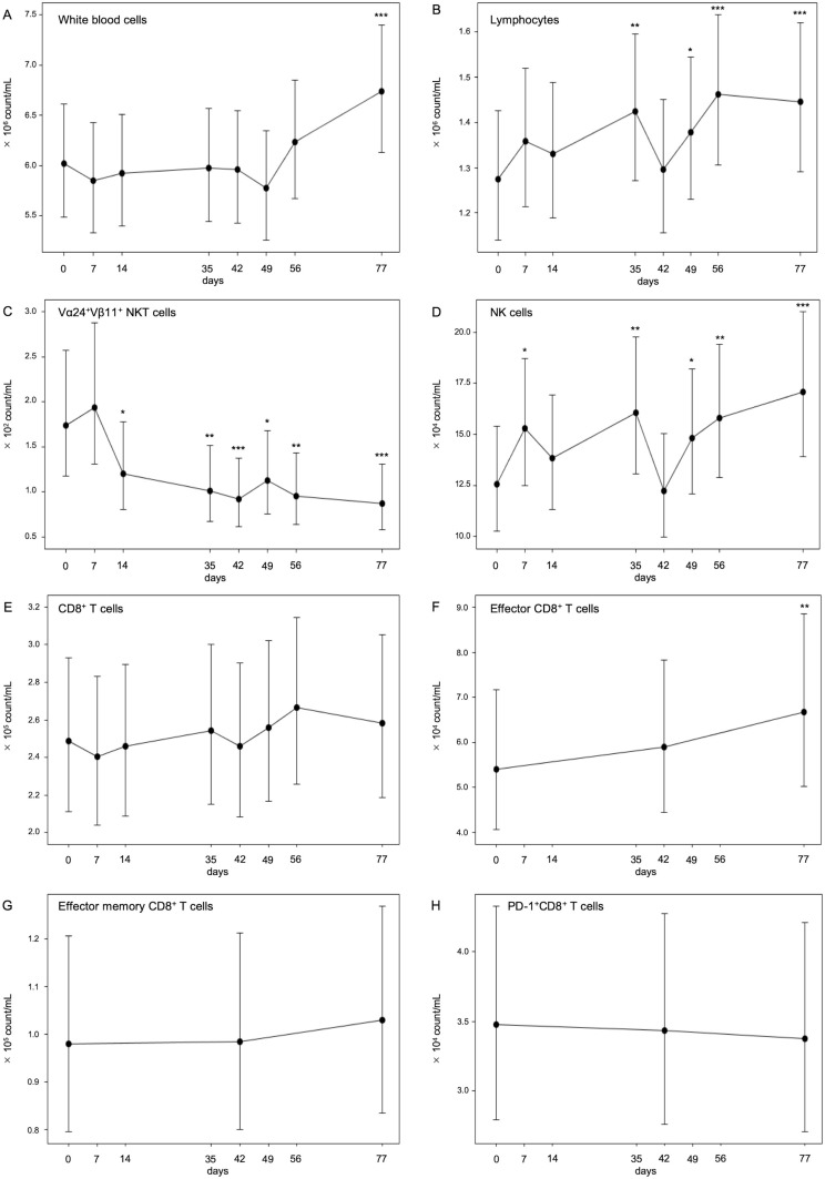
Figure 3.
Numbers of circulating lymphocytes during treatment. The absolute numbers of white blood cells (A) and lymphocytes (B) were counted by clinical blood tests. The percentages of peripheral blood Vα24+Vβ11+ iNKT cells (C), CD56+CD3− NK cells (D), CD3+CD8+ T cells (E), CD45RA−CCR7+ effector CD8+ T cells (F), CD45RA−CCR7− effector memory CD8+ T cells (G), and PD-1+CD8+ cells (H) during treatment were assessed by flow cytometry analysis, and the absolute numbers of these cells were calculated using full blood counts. The linear mixed effect model for repeated measurements was used to estimate the geometric mean, SE, and 95% CI of the mean of each measuring point of immune monitoring, which can address all available post-baseline data. *p<0.05, **p<0.01, ***p<0.001. NK, natural killer.

