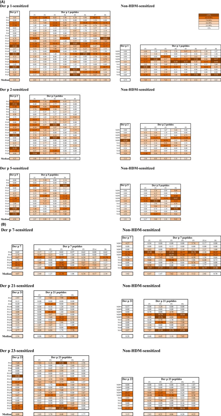
Figure 6.

T cell reactivity of Der p allergens and allergen‐derived peptides for each subject. Shown are percentages of proliferated CD4+ T cell in responses to the allergens and allergen‐derived peptides (A, Der p 1, Der p 2, Der p 5, B, Der p 7, Der p 21, Der p 23; Means of triplicates; color codes for different percentages of proliferated cells are shown with a cut off of 2%‐white) in PBMC cultures from each allergen‐sensitized patient (PA), non‐HDM‐sensitized allergic individuals (NDP) and nonallergic individuals (NA). Median percentages are shown for each allergen and peptide for sensitized and nonsensitized individuals at the bottom of each Table
