
Figure 3.
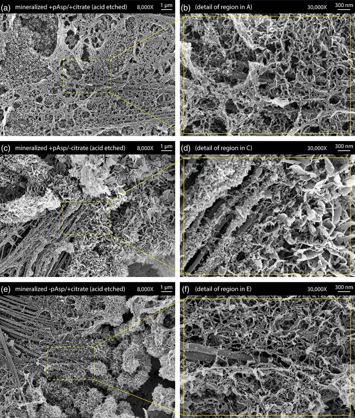
Scanning electron microscopy (SEM) images of acid‐etched mineralized collagen sheets. (a,b) Sheets prepared with pAsp and citrate additions to the mineralization treatment process showed mineral bridging between collagen fibrils. (c,d) Absence of citrate from the mineralization process led to extrafibrillar plate‐like crystals growing much larger in size. (e,f) Absence of pAsp from the mineralization process led to excessive accumulation of extrafibrillar spherulites