
Figure 4.
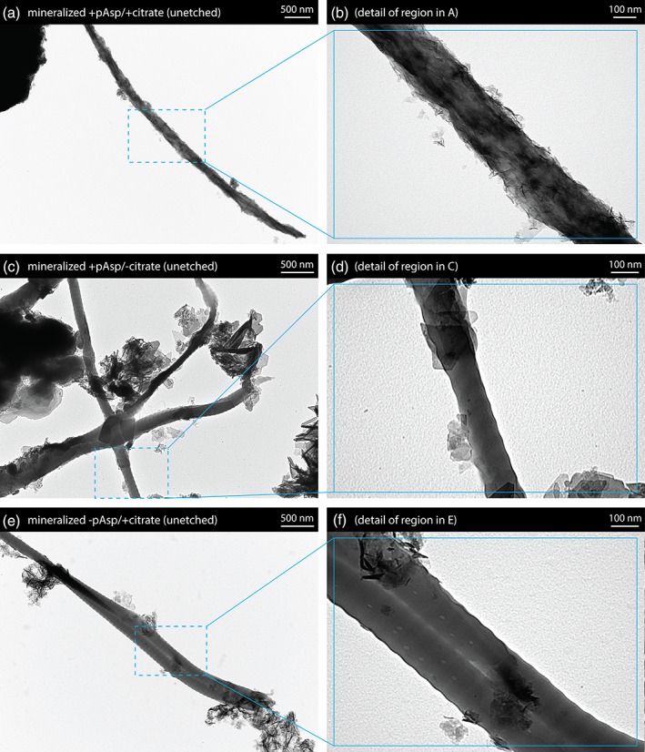
Transmission electron microscopy (TEM) images of collagen fibrils removed from the unetched mineralized collagen sheets. (a,b) Sheets mineralized with pAsp and citrate additions contained collagen fibrils incorporating a high density of small, longitudinally aligned crystals. D‐banding was not apparent, presumably due to the quantity of mineral present. (c,d) For sheets mineralized without citrate, much larger plate‐like crystals were seen. (e,f) Sheets prepared without addition of the PILP process directing agent pAsp contained fibrils with a reduced quantity of closely associated mineral