
Figure 1.
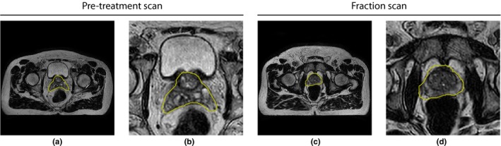
Examples of pretreatment (a) and daily fraction scans (c) with ground truth prostate contours from the same patient in yellow. Both images show the center slice of the volume. (b) and (d) show zoomed‐in versions of (a) and (c), respectively. The images are from T2‐weighted three‐dimensional (3D) turbo spin‐echo scans. [Color figure can be viewed at http://wileyonlinelibrary.com]