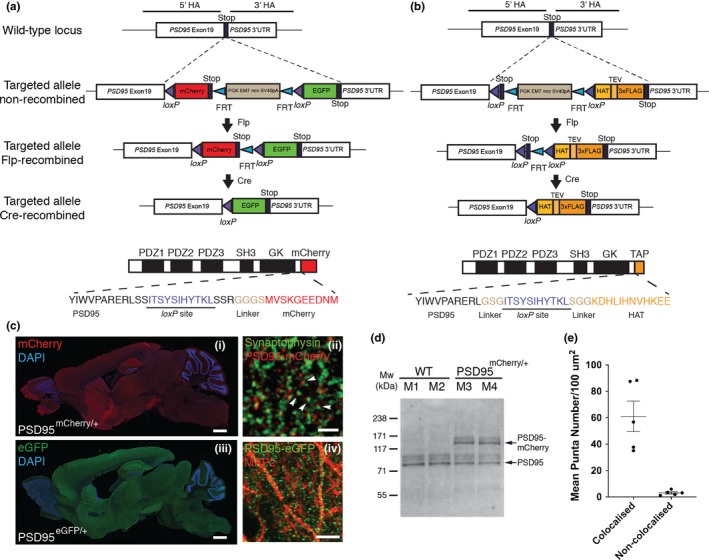
Figure 1.

Generation of PSD95c(mCherry/eGFP) and PSD95cTAP knock‐in mice. (a) Gene targeting strategy for the PSD95c(mCherry/eGFP) mice. The PSD95 (Dlg4) allele was targeted with a tandem fluorescent tag (mCherry and eGFP) coding sequence inserted at the last exon and immediately before the STOP codon. The FRT site‐flanked neo gene was removed by crossing PSD95c(mCherry/eGFP) mice with FLPe deleter mice. The progeny PSD95c(mCherry/eGFP) (without neo) mice were further bred with different Cre driver lines. Bottom panel shows domain structure of PSD95‐mCherry fusion protein, which contains three PDZ, an SH3, a GK domain and the C‐terminal mCherry tag (before Cre recombination). Note that a short peptide encoded by the loxP site and a linker sequence were inserted into the open reading frame of PSD95. (b) Gene targeting strategy for the PSD95cTAP mice. By a similar targeting strategy, a loxP site‐flanked STOP codon and the TAP sequence were inserted before the PSD95 STOP codon. Bottom panel shows the domain structure of PSD95‐cTAP fusion protein (after Cre recombination), which includes the C‐terminal‐tagged TAP tag. (c) Ubiquitous PSD95‐mCherry/eGFP expression in adult mouse brain before (i) and after (iii) breeding with a germline CAG‐Cre driver line. Note that both PSD95‐mCherry (identified by anti‐mCherry antibody immunostaining, i) and PSD95‐eGFP (identified by native eGFP fluorescence, iii) are widely expressed across the brain and show a similar distribution pattern. Scale bar: 0.5 mm. (ii) Fluorescence confocal image of brain sections of fluorescent knock‐in PSD95mCherry/+ mice; PSD95‐mCherry puncta (red) are located in close opposition to the anti‐Synaptophysin‐stained pre‐synaptic terminals (green; arrowheads). Scale bar: 2 μm. (iv) Representative image of anti‐MAP2 immunofluorescence staining on PSD95eGFP/+ brain sections. Discrete PSD95‐eGFP puncta (green) were detected along the MAP2‐staining neuronal processes. Scale bar: 10 μm. (d) Western blotting analysis of homogenate extracts from wild‐type (M1, M2) and littermate heterozygous (M3, M4) PSD95mCherry/+ mice, using antibodies against murine PSD95. (e) Mean punctum number/100 µm2 shows that the majority of PSD95‐mCherry puncta are in close opposition to (defined as “colocalisation”) Synaptophysin‐labelled pre‐synaptic terminals. PSD95‐mCherry and Synaptophysin‐positive puncta were manually quantified using ImageJ plugin Cell Counter (Kurt De Vos). Error bars: mean ± SEM; unpaired, two‐tailed t test, p = .0011, n = 5 cryosections [Colour figure can be viewed at http://wileyonlinelibrary.com]
