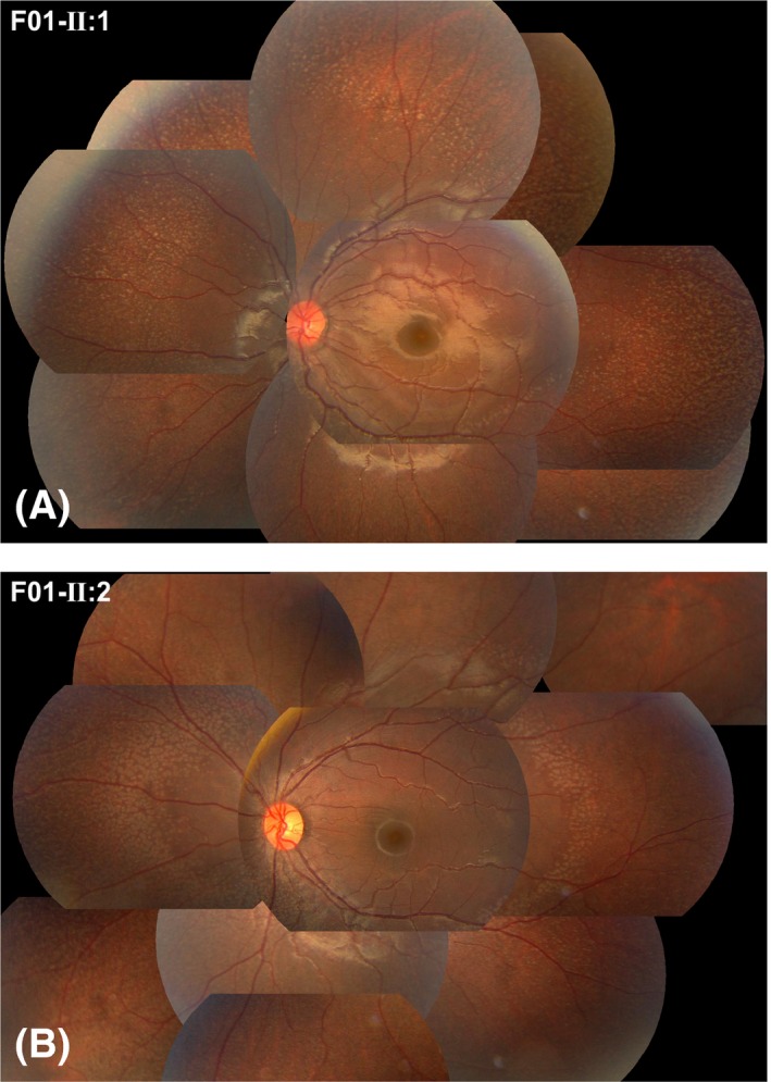
Figure 2.

Photos demonstrating the fundus changes typical of fundus albipunctatus‐like changes in two affected brothers in family F01. A number of grey‐white dots were present in the mid‐peripheral retina. The family number and individual ID number that correspond to those shown in Fig. 1 and Table 2 are listed on the top left corner of each photo, as shown in Figs 3, 4, 5.
