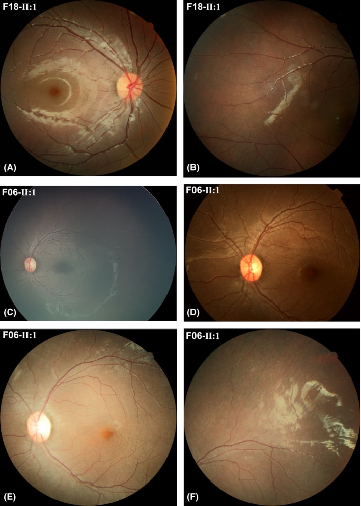
Figure 5.

Mild fundus change and progression in retinal degeneration. (A, B) Fundus photos obtained from F18‐II:1, who had high hyperopia. The photos were taken when the patient was 6 years and 3 months old and showed a normal‐like posterior fundus and mild degenerative changes at the mid‐peripheral retina. He had a corrected visual acuity of 1.0 in the right eye and 0.5 in the left eye. (C–F) Fundus photos obtained from F06‐II:1. The photos were taken when the patient was 2 years and 2 months old (C), 5 years old (D) and 8 years and 10 months old (E, F). The fundus changes were insignificant or very minor in the early stage (C, D) but an obvious tapetoretinal degeneration was observed in the posterior (E) and mid‐peripheral retina (F) when the patient was 8 years and 10 months old.
