Fig. 2.
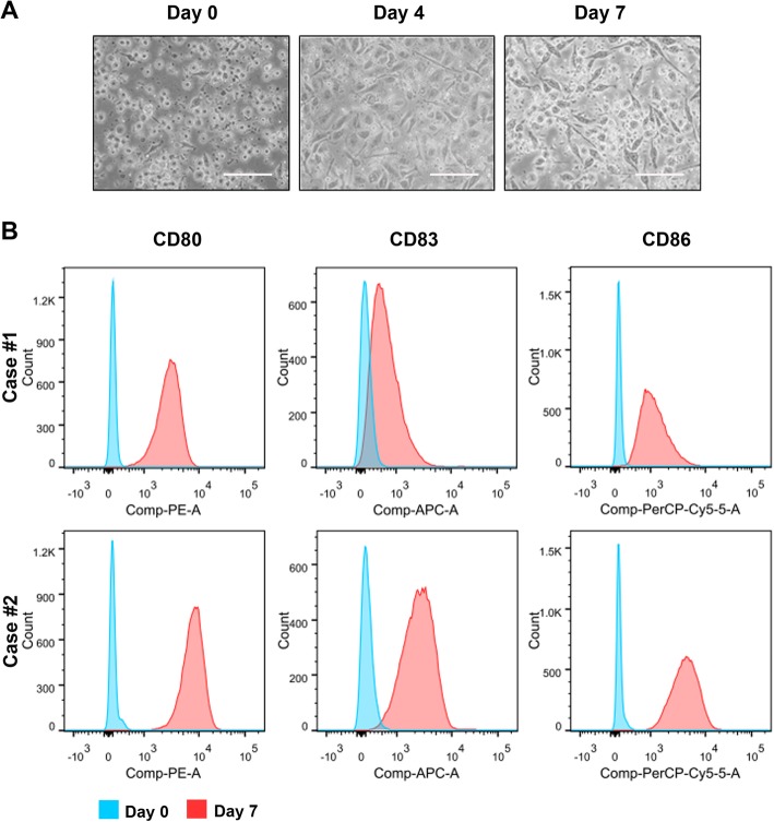
Phenotypic characterization of DC cells. a Representative images of adherent cultured PBMC-isolated precursor monocytes (day 0), inflammatory cytokine-stimulated maturing DC cells (day 4), and peptide-loaded mature DC cells (day 7). The peptide-sensitized DC cells exhibited the typical dendritic appearance. Magnification, × 40; bars, 200 μm. b FACS analysis. Results showed that the peptide-stimulated DC cells, isolated from two representative PBMC preparations, expressed higher levels of three DC-associated surface markers, CD80, CD83, and CD86

