Fig. 2.

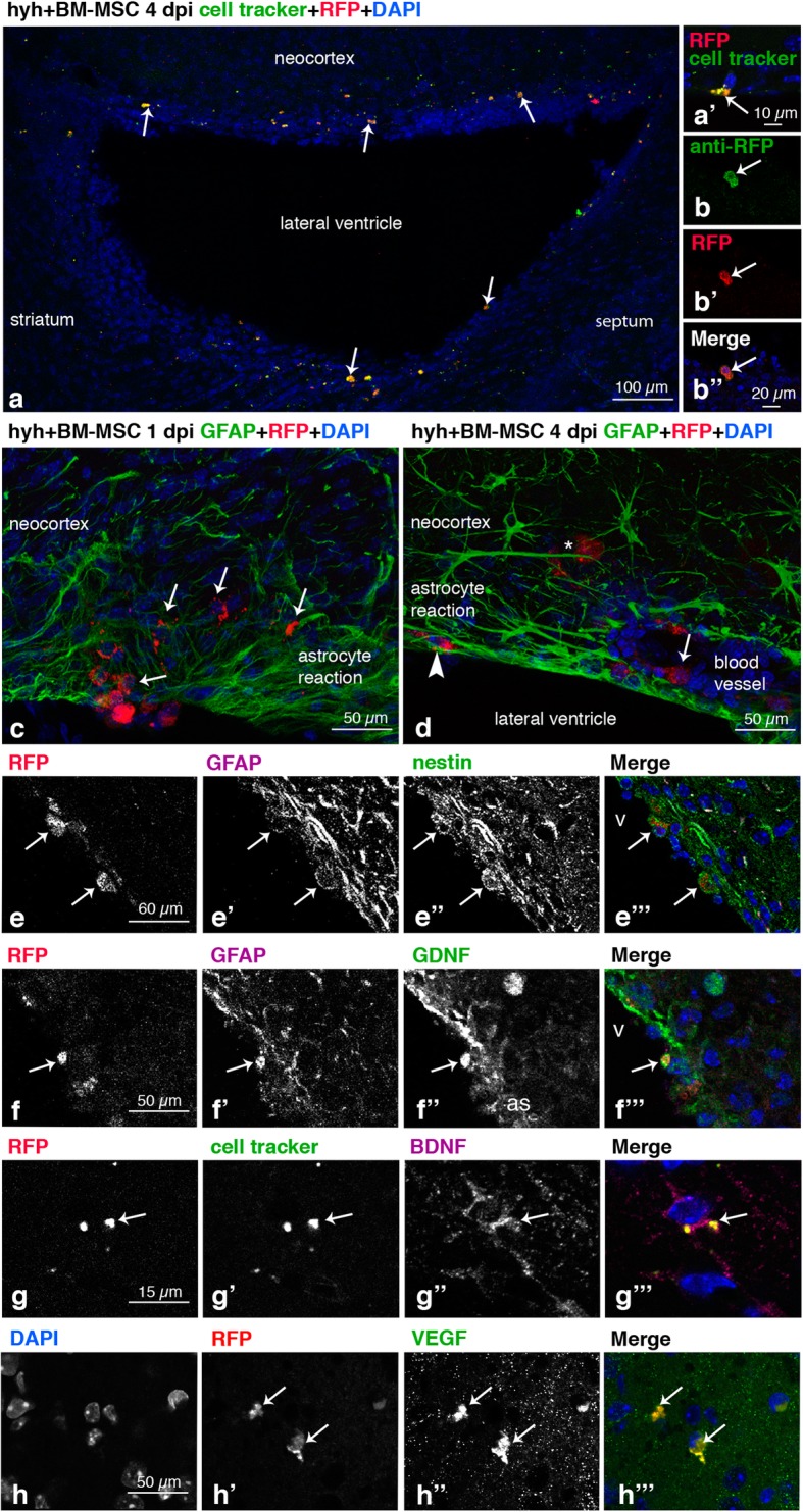
Location of BM-MSC in the hosting tissue and detection of neuroprotector factors expression. a Walls of the lateral ventricle of a hyh mouse administered at 20 days of age with BM-MSC expressing the mRFP1 (red) and labeled with a green cell tracker (white arrows), 4 days post-injection (dpi). a’ Detail of a BM-MSC (RFP fluorescence, red) colabeled with the fluorescent green cell tracker (white arrow). b, b” Colabeling of the mRFP1 (red) with an antibody against RFP (green) in the administered BM-MSC, 4 dpi, in the neocortex of a hyh mouse injected at 20 days of age. c BM-MSC (red, white arrows) entering into the brain parenchyma of a hyh mouse 20 days of age, 1 dpi, through a ventricle surface presenting a loose periventricular layer of reactive astrocytes (GFAP immunolabeling, green) in the neocortex wall. d In the neocortex walls of hyh injected at P20, BM-MSC were found in three different locations at 4 dpi: between the dense layer of reactive astrocytes covering the ventricle surface (arrowhead, GFAP immunolabeling in green), around the blood vessels (arrow), and deep into the brain parenchyma (asterisk). e–e”’ Coexpression in BM-MSC (mRFP1, red; arrows) at 4 dpi in the neocortex wall of GFAP (magenta) and nestin (green). f–f”’ Coexpression at 4 dpi in BM-MSC (red mRFP1, arrow) of GFAP (magenta) and GDNF (green). g–g”’ BM-MSC (mRFP1, red; arrow) colabeled with the green cell tracker and anti-BDNF (magenta) at 4 dpi. h–h”’ Expression in BM-MSC (red, mRFP1; arrows) of VEGF (green) at 4 dpi
