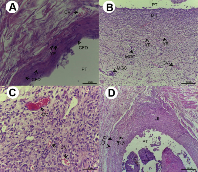
Fig. 2.

( A ) Fifteen days after implantation, Group C: presence of congested vessels (CV), young fibroblasts (YF), and collagen fibers disposed (CFD) in parallel bundles involving the cavity ( HE, 400×magnification, scale: 25 µm). Area of polyethylene tube (PT) implant. ( B ) Fifteen days after implantation, Group M10: cavity surrounded by moderate inflammatory infiltrate (IIM), CV, presence of ovoid and fusiform YF, and multinucleated giant cells (MGC) (HE,100×magnification, scale:100µm). Area of PT implant. ( C ) Fifteen days after implantation, Group KC10; moderate inflammatory infiltrate with intense vascularization with CV of various sizes (HE, 400× magnification, scale: 25 µm). ( D ) Fifteen days after implantation, Group KC50: deposition of delicate CFD in parallel bundles, ovoid and fusiform YF, and light inflammatory infiltrate (LII) (HE, 100× magnification, scale:100 µm). Area of PT implant. HE, hematoxylin and eosin.
