
Fig. 3.
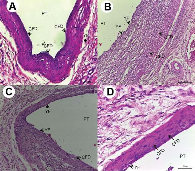
( A ) Thirty days after implantation, Group C: thick layer of collagen fibers disposed (CFD) in parallel bundles in the midst of scarce young fibroblasts (YF) (HE, 200× magnification, scale: 50 µm). Area of polyethylene tube (PT) implant. ( B ) Thirty days after implantation, Group M10: cavity surrounded by deposition of CFD in parallel bundles and YF (HE, 100× magnification, scale:100 µm). Area of PT implant. ( C ) Thirty days after implantation, Group KC10: deposition of CFD in the midst of ovoid and fusiform YF (HE,100× magnification, scale:100µm). Area of PT implant. ( D ) 30 days after implantation, Group KC50: layer of CFD in the midst of dispersed ovoid and fusiform YF (HE, 400×magnification, scale:25 µm). Area of PT implant.