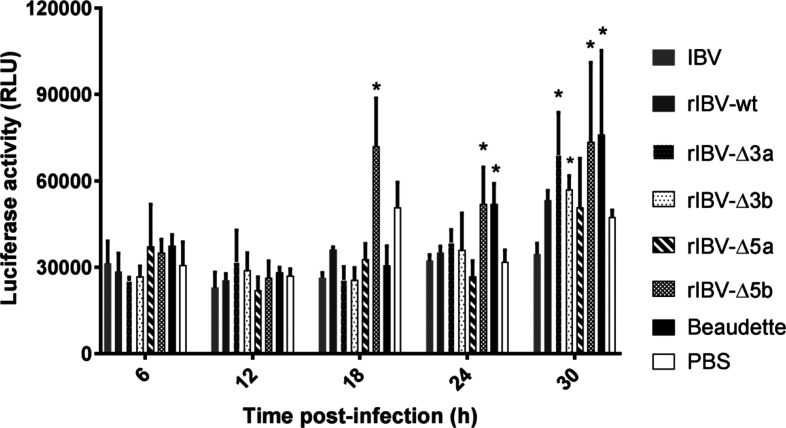
Fig. 5.
CEK cells were infected with Beaudette, IBV H52 BI, rIBV-wt, rIBV-Δ3a, rIBV-Δ3b, rIBV-Δ5a and rIBV-Δ5b at a multiplicity of infection (m.o.i.) of 10 in triplicate. Culture supernatants were collected at five time points and used to infect CEC-32 cells in duplicates. The values represent the means of six measurements for each virus at each time point, and the bars indicate sd. *, significant differences (P<0.05) as compared to wild-type H52 viruses.

