Fig. 3.
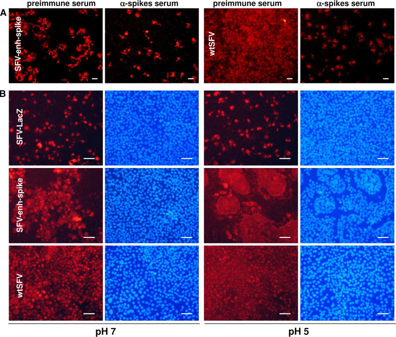
Propagation of SFV without capsid is mediated by the envelope proteins and does not require cell fusion. a BHK cell monolayers were infected with SFV-enh-spike VPs at MOI 0.1, or with wtSFV at MOI 0.025, and incubated for 24 h (SFV-enh-spike) or 10 h (wtSFV) in the presence of α-spikes serum, or with preimmune serum (in both cases diluted 1:50). b BHK cells were infected with SFV-enh-spike and SFV-LacZ VPs at MOI 0.05, or with wtSFV at MOI 0.025, and 16 h later were incubated during 3 min with PBS at the indicated pH. Cells were washed and incubated for 3 h with BHK complete medium. In both a and b, cells were fixed and analysed by IF with α-spikes (a) or α-nsp2 sera (left images for each pH in b). Nuclei in (b) were visualized with DAPI (right images for each pH). Magnification: ×100 (a) or ×200 (b); scale bars 50 μm. Panels correspond to a representative experiment from at least two independent experiments performed in triplicate with similar results. See also Supplementary Fig. 3 for higher magnification images in which cell membranes have also been labelled

