
Fig. 6.
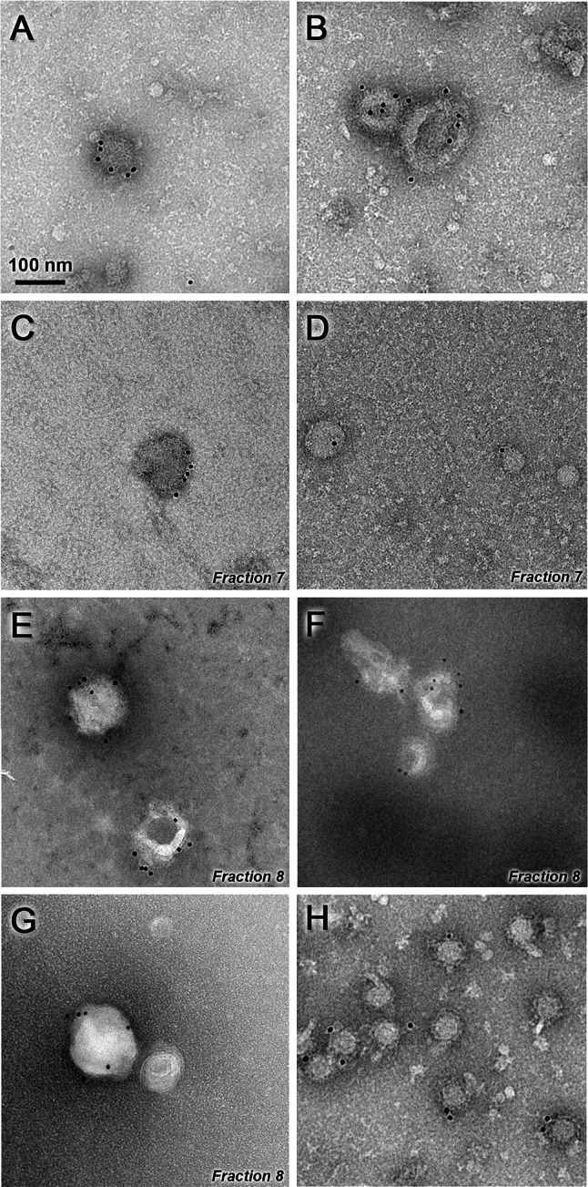
Electron microscopy of iMVs. Purified iMVs were incubated with an antiserum specific for SFV envelope proteins, followed by incubation with a gold-labelled secondary antibody and visualized by negative staining (a–g). Representative images of iMVs present before sucrose gradient ultracentrifugation (a, b) or in the indicated fractions after gradient purification (c–g) are shown. SFV-LacZ VPs isolated as described in “Materials and methods” were used as positive control (h). Magnification in all pictures is ×30,000. Scale bar 100 nm