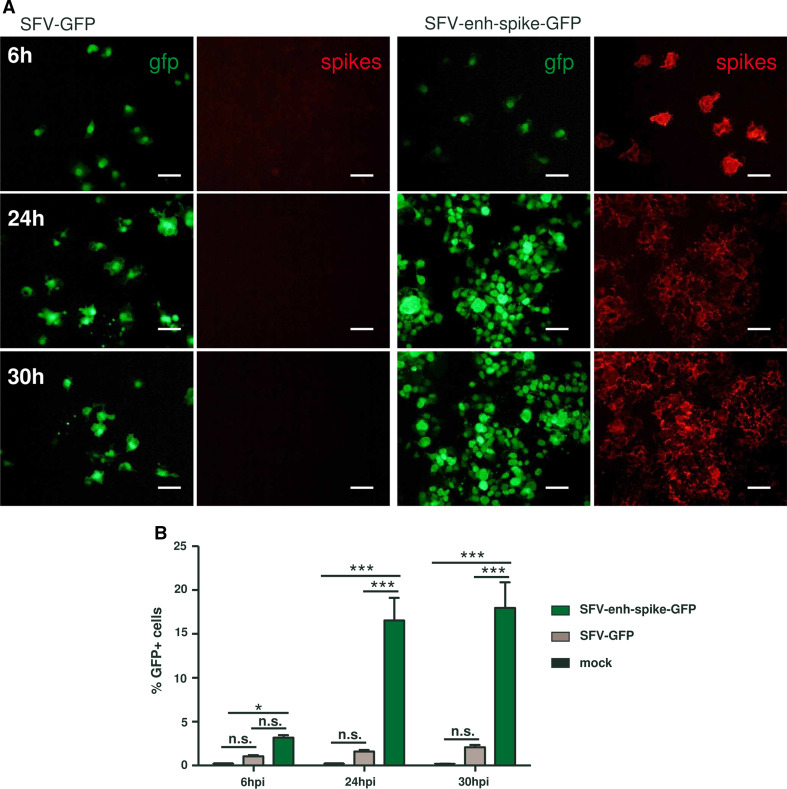
Fig. 8.
Propagation of SFV-enh-spike-GFP. BHK cell monolayers were infected with the indicated vectors at MOI 0.5 (a) or MOI 0.05 (b). a Cell monolayers were fixed at the indicated times and GFP expression was visualized with the appropriate filter. Spike expression was analysed in the same preparations by indirect immunofluorescence with an antiserum specific for SFV envelope proteins. Magnification of images: ×200; scale bars 50 μm. b Cells were trypsinized, fixed with 2 % paraformaldehyde and analysed by flow cytometry (FACS-Canto II, BD-Biosciences) to determine the percentage of GFP-positive cells. Data analysis was carried out using FlowJo software (Tree Star Inc., Ashland, OR). Results correspond to one of two representative experiments, each performed in triplicate that gave very similar results. Error bars represent the mean + SD. *p < 0.05; ***p < 0.001; ns not significant

